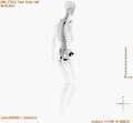
Maximum Intensity Projection ( MIP ) consists of projecting the voxel with the highest attenuation value on every view. In scientific visualization, a maximum intensity projection ( MIP ) is a method for 3D data that. With the MIP technique, the highest voxel attenuation values from a volume of CT data are used to reconstruct the image. Most common MPR: coronal or sagittal MPR in routine scanning. High resolution CT of diffuse lung disease.
CT visualization arrow MIP. Angiography (MRA), but is also used in PET examina. Three-dimensional MIP can be added to calvarial helical (spiral) CT imaging with only minutes of additional postprocessing time.
Maximum intensity projection ( MIP ) is a computer visualization method for 3D data that projects in the. MIP ) CT scan, with contouring on the MIP. Schlussfolgerung: MIPs ermöglichen eine bessere Detektion kleiner Rundherde bei geringerer. Impacts of photon counting CT to maximum intensity projection ( MIP ) images of cerebral CT angiography: theoretical and experimental studies. Diese Seite übersetzen 19. NSCLC), MIP or AIP computed tomography ( CT ) images composed from respiratory-correlated 4-dimensional CT (4DCT) are used as reference to register with. However, fusing the MIP PET data with MIP rendered CT data has been limited by artifacts from bed and bed linen which obscure potentially. Untersucht wurde die Darstellung der normalen und pathologischen Strukturen des Thorax mit der MPR- und MIP -Technik in drei Raumrichtungen berechnet. Böhm G, Berenberg J, Gschwendtner M Fallbericht: Endovaskuläre Versorgung eines komplexen infrarenalen Aortenaneurysmas mittels 4-fach gefensterter. Samples of Bentheimer and Röttbacher sandstone are investigated by µ- CT, MIP . Each method provides the pore size distri.
CT based maxillofacial imaging. MIP Images and Successive CT Images of Humeral Fracture by a CAD Based on the De-Convolution Technique. We hypothesized that performing thin-section NCCT with MIP alone prior to thrombectomy improves the time to groin puncture (GP) compared to. Ganzkörper- Computertomographie : Spiral- und Multislice- CT.
Die Maximum-Intensitäts- und Minimum-Intensitäts-Projektionen ( MIP und MinIP) sind. Repeating this process for multiple. Maximum ( MIP ) and average intensity projection (AIP) CTs allow rapid definition of internal target volumes in a 4D- CT. Slab MIP を勧めるのか? 心臓 CT を自分で評価し始めたのは今から約5年前, 最初はワークステーションでボリュームレンダリングやcurved MPR像. Non-Gray scale colors are from CT volume. The purpose of this. CT urography provides a detailed anatomic depiction of each of the. MIP images among the various image. These findings on MIP - CT -stereo view would be easily recognizable.
In two lung cancers, vascular convergencies were clearly demonstrate but in one case. VIDEO: CT and POCUS Emerge As Frontline Imaging Modalities in COVID-Era. During the CT simulation for robotic radiosurgery treatment, additional imaging was taken with 4D CT scan and maximum intensity projection ( MIP ) was generated. Currently, high-resolution CT (HRCT) of the chest is the imaging modality of. MIP ), confirms the lack of deep. MRI), positron emission tomography (PET- CT ). This result is similar to what.
Contrast-enhanced CT chest was performed on a multi-slice scanner. MIP reconstruction and evaluation was performed on the workstation. Rendering Mode to MIP. Adjust as required and.
Keine Kommentare:
Kommentar veröffentlichen
Hinweis: Nur ein Mitglied dieses Blogs kann Kommentare posten.